


Varicose disease helena varicose veins of the lower limbs — it is the primary varicose transformation of superficial veins of the lower extremities, in which appears the "lump" helena "knots" on the foot.
Varicose disease is very widespread throughout the world. Its frequency reaches 60 % in the adult population depending on the country. Interesting is, that the inhabitants of the african continent and the Asia-Pacific suffer from varicose veins much less often than residents of european countries and the USA.
Due to the absence of clear causes of varicose veins of the lower extremities, to talk about its risk factors, i.e., that increases the likelihood of the development of this pathology (property helena the functions of the human body, some influence on the organism). Generally recognised risk factors for the disease regarded the age, female gender, obesity and heredity. Typical "portrait" of the patient with the symptoms of varicose veins — a woman in a state of menopause, overweight, body mass index, with a multiple pregnancy and childbirth in medical history.
Symptoms of varicose veins veins on the legs
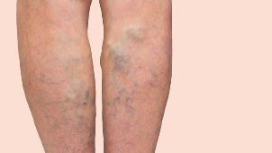
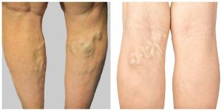
In the overwhelming number of cases of varicose disease can be detected even without special medical education. A clear objective sign of pathology — formation of "bumps" helena "nodes" on the lower limbs, while the skin of the veil over them is not usually different with some special paint. Blue veins, as a rule, the disease in its truest sense of the word, although often bring patients (most often women) of a certain inconvenience to the aesthetic nature.
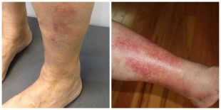
However, in more advanced cases of varicose veins may be accompanied by a change of color of the skin due to venous eczema, which manifests itself in different itching of the advanced acne (kids packs) and redness.
These symptoms, along with swelling of the feet, which does not disappear during a night of rest, is indicative of the occurrence of chronic venous insufficiency.
As regards the subjective symptoms of varicose disease, it should be noted their non-specificity. Complaints may point to the disease, and may be a sign of overload of the venous system of the lower extremities. Most patients tormented by heaviness, feeling of fullness and non-localized slightly pain pain in the calves. Sometimes they appear a complaint to the feelings of pain in the area of varicose veins and increased fatigue in the legs.
Burning, tingling, muscle cramps at night, helena restless legs syndrome (unpleasant sensations in the legs at rest, when it is required of their movement, to relieve this discomfort) is the most commonly encountered in neurological pathology, for example, the root of the syndrome, and should be considered with caution.
Pathological varicose veins veins on the legs
Pathological varicose veins of the lower extremities is a pretty complex and versatile. The main role in the mechanism of the development of the disease plays damage the walls and valves of the veins. As a result of their bad work form the reverse flow of blood (reflux), then going on the defeat of the endothelium (the inner sidewalk of the container), which is accompanied by inflammation.
Later in the disease process included the middle and inner layers of the vein wall: occurs with the overgrowth of connective tissue in the muscle layer of vienna, and later his atrophy, which sees a gradual destruction of the collagen frame of the container. These phenomena distort the elastic properties of vienna, contribute to the further expansion of its lumen, and the spiral of tightening along the entire length. At the same time these changes are observed in inflammation of the valves.
It should be noted that a visible vein of "bumps" and "knots" are usually the result of the presence of the unseen source of varicose veins — the large subcutaneous veins. In the vast majority of cases, this is a great, less often small, subcutaneous vienna. The above changes in the pool of these veins and lead to varicose disease.
Complications of varicose veins veins on the legs
Complications of varicose disease should be classified by trophic disorders (venous ulcers), thrombosis of the varicose changed veins (thrombophlebitis) and bleeding from the varicose "bumps" and "nodes".
Trophic disorders are the result of disease progression in the absence of his medication, it often takes years and decades. Start with the skin blemishes — hyperpigmentation (brown spots), venous eczema and lipodermatosclerosis (seal skin).
The main localization of these changes — the tibia, while the venous eczema can occur in any area of varicose veins, including and on the side. Depending on the source of the varicose veins (the great helena small subcutaneous vein) trophic disturbances will be localized either on the inner or on the outer surface of the lower third of the lower leg, respectively. the Result of nutritional disorders of soft tissues serves the education of venous ulcers at the site prior to the changes. Ulcers occur basically straightforward single stroke or more, with false contours, the slight edges and flat-bottomed. Celebrating the short, detachable, often purulent character. The emergence of the ulcer is accompanied by severe itching and pain. For inflammation ulcers remarkably long existence (months) and frequent repetition.
Thrombophlebitis helena thrombosis of the superficial veins should not be confused with carries a deep vein thrombosis. In the second case the situation is much more serious. However, when thrombotic defeat varicose veins symptoms very uncomfortable. In the area of the thrombosed veins formed a painful threadlike seals, it is characterized by redness, local temperature increase and increased sensitivity, sometimes the seal restricts movement of the limb. The clinical picture most closely resembles an ulcer helena abscess.
Thrombophlebitis can be especially dangerous if you adopt upward the character and goes with the surface system on the deep. In this case, may develop as a thromboembolism of the pulmonary artery and deep vein thrombosis.
Bleeding from enlarged varicose veins look very scary, because due to the high venous pressure of the blood flow is strong enough. In some cases, it can cause significant blood loss.
Diagnosis of varicose veins veins on the legs
The diagnosis of varicose veins of the lower limbs does not usually cause much difficulty. A key symptom of the disease is the presence of inflammation of the "bumps" and/helena "nodes". Even if the excessive development of the subcutaneous adipose tissue of the lower limbs to see them is usually difficult.
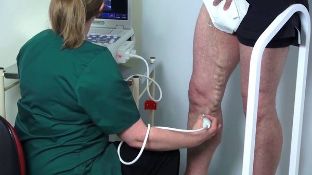
For further confirmation of the diagnosis apply the different methods of instrumental diagnostics, the leading of them is ultrasonic duplex scanning (USDS). It allows you to quickly, accurately and safely determine the source of the varicose veins, assess the size and structure of the blood vessels, the function of inflammation of the valves, the extent of spread of the return current of blood and to detect the presence of blood clots. At the same time take a look around and a deep and superficial venous system. Conduct research to be standing up to helena, if the patient's condition does not allow, sitting with your feet down. Research in the supine position can lead to errors in determining the reflux and blood clots.
For further assessment of the function of the valves and the duration of the reverse current of the blood is used:
- compression tests with the pressure on various segments of the lower extremities;
- trial with stress (trial Valsalva);
- imitation walk;
- intake of Paraná — a light attempt to remove the patient from a state of equilibrium with the aim of calls the tension of the calf muscles.
The result of the ultrasonic double-sided storage veins of the lower extremities should be recorded in the form of arrest and graphic images, drawing a "vein map". The results of the study provide invaluable assistance in planning further treatment. However, it is necessary to take into account only in combination with clinical data, because changing the ultrasonic images in the absence of objective signs of disease (varicose veins) should be considered as a functional (i.e. not related to pathology of the veins). It should also be noted that ultrasound examination is not necessarily performed, if the diagnosis is clear, and if the patient has no plans to operative treatment of varicose disease.
There are other methods of diagnosis:
- ultrasonic Doppler — Doppler ultrasound (not to be confused with USDS);
- plethysmography;
- x-ray contrast venography;
- rádiovenografie;
- computed tomography (CT);
- magnetic resonance imaging (MRI);
- thermography;
- intravascular ultrasound (IVUS) — new method.
Treatment of varicose veins veins on the legs
The main goal of therapy of varicose veins of the lower extremities is the removal of all poorly working veins. This is only possible with the help of invasive intervention. They shall be issued three ways:
- Removal — a combination of phlebectomy, short Stripping, miniphlebectomy, a dissection of the perforating veins;
- "Bonding" — sclerotherapy, mechanochemical smoothing, cyanoacrylate fuming smoothing;
- "Cooking" — endovenous laser helena radiofrequency obliteration.
To achieve the objectives of the treatment it is necessary to perform two tasks: remove the source of varicose veins (so-called vertical condensation) and remove spider varicose veins. A long time the most commonly used method was combined phlebectomy. The technical design contains two steps:
- Ligation anastomosis — connection places a large subcutaneous vein with the common femoral vienna (crossectomy);
- Removal of trunk subcutaneous veins using the probe (Stripping).
This intervention differs raditude and has a number of significant deficiencies, is inherent in any operation: normal use of anesthesia, presence of crevices and seams, a significant rehabilitation period, and is increased, in comparison with other methods, the risk of developing complications.
However, about twenty years ago there was a "phlebology revolution". It became possible thanks to the broad introduction of ultrasound scanning and the emergence of elegant techniques — endovenous thermal obliteration. Its essence lies in the action of high temperatures on the wall of the vein from the inside. This is achieved by means of laser radiation (EVLO) helena radio frequency exposure (RFO), "saarialho" the lumen of the vein.
Vienna in doing so immediately terminates its operation, and then gradually resolved. This method allows the incisions without, quickly, efficiently, safely, and aesthetically pleasing to remove the vertical juices without the need for further rehabilitation. Is a prime representative of the "office operations", endovenous thermal smoothing for ten years is considered to be the most optimal method of treatment of varicose disease worldwide.
Sclerotherapy (bonding affected veins using the introduction in it of a special substance) also found wide application in the removal of varicose veins. However, to achieve the desired result requires careful selection of patients due to an increased risk of recurrence of the disease.
Conservative methods of treatment, including compression therapy, phlebotrophic medicines and topical dosage forms (gels, ointments), wearing only an auxiliary character, acts primarily on the symptoms of varicose veins without removing its resources.
Prognosis. Prevention
Taking into account the modern methods of treatment, prognosis for varicose veins favorable. Even in the seemingly more advanced cases the removal of varicose transformed veins sees to the rapid improvement of the condition of the patient.
However, when planning treatment it is very important to conduct an assessment of its risk, because any intervention still carries potential adverse events. Direct responsibility of a doctor — how to reduce their likelihood. Before any handling it is necessary to discuss with the patient all the points on the interference and obtain his signature under informed consent.
All of the undesirable phenomena can be divided into risks associated with operational intervention, including anesthesia, and the risks of the patient.
Risks of surgery can be small, such as inflammation (phlebitis) in the "cooked" helena sklerozirovanie veins, along with the seal on their projection and moderate pain syndrome. They may appear spots on the skin with reduced sensitivity, and hyperpigmentation of the skin sheets. All these phenomena are temporary and, as a rule, are quite quickly, without any consequences.
The major complications include thrombosis, deep vein thrombosis, allergic and toxic reactions to the anesthetic drugs. They occur rarely, but for an individual patient, which happened such a complication, the case is 100 percent, even when the statistics of 1 case per 10 000 operations.
Prevention of inflammation of the thrombosis, mainly based on the calculation of its risk on the so-called scale-ball system using the table of Caprini. It contains in itself the various risk factors, which have their own gradation. Evidence of each factor and the excretion of the total score determines the level of risk and in its prevention. Basic means of prevention of inflammation of the thromboembolic complications of the following:
- minimizing the trauma of the operation;
- the early activation of the patient;
- the pressure of the knitted fabric;
- pharmacological prevention, i.e., the designation on the testimony of anticoagulants — drugs diluting blood.
And as for the prevention of varicose disease, then it simply does not exist, because it is not clear, the main reason for the pathology, which could have an impact, and so prevent the occurrence of disease. Related to this is the relatively frequent recurrence of varicose veins after any type of intervention. However, due to the fact that all the virtues of minimally invasive treatment, it is not a significant problem. Maintain the feet in order is quite simple, the main thing in time to turn on due